3-Color IHC Kit for 1 Mouse/2 Rabbit Antibody on Rodent Tissue, Brown/Red/Geen

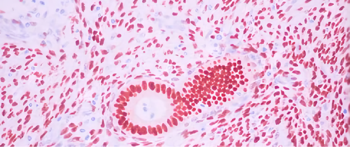
Triple staining employs both conventional and innovative techniques in immunohistostaining to unveil the presence of three separate antigens and their simultaneous expression within a single tissue. The 3-Color IF Kit is designed to use DAB(brown), Permanent Red (red), and Emerald (green) dye effectively stain 3 different antigens on rodent tissue or cell samples when paired with user-supplied 1 mouse and 2 rabbit primary antibodies.
Information
Kit Contents
HRP-Polymer anti-Rabbit (RTU)
HRP-Polymer anti-Mouse (RTU)
AP-Polymer anti-Rabbit (RTU)
Mouse Primer (RTU)
DAB Substrate (RTU)
DAB Chromogen (20x)
Permanent Red Substrate (RTU)
Permanent Red Activator (5x)
Permanent Red Chromogen (100x)
Antibody Blocker (40x)
Mouse-Rabbit-Rabbit Blocker 1 (RTU)
Mouse-Rabbit-Rabbit Blocker 2 (RTU)
Emerald Chromogen (RTU)
Mounting Medium (RTU)
Usage
The kit supplies polymer enzyme conjugates, namely polymer-HRP anti-mouse IgG, polymer-HRP anti-rabbit IgG, and polymer-AP anti-rabbit IgG, accompanied by three substrates/chromogens: DAB (brown), Emerald (green), and Permanent Red (red). It utilizes a non-biotin system to prevent non-specific binding caused by endogenous biotin. This kit has been optimized to eliminate cross-detection when detecting more than two primary antibodies from the same host species using a unique blocking system.
Applications
Paraffin tissue (verified), frozen specimen and freshly prepared monolayer cell smears.
Color
DAB (brown),Permanentred (red),and Emerald (green)
Specificity
Mouse and Rabbit
Tissue Species
Rodent
Storage
2-8℃
Note
The outcome is significantly influenced by several factors, including fixation, tissue slide thickness, antigen retrieval, as well as the dilution and incubation time of the primary antibody. It is crucial for the investigator to carefully evaluate all of these variables and establish the ideal conditions in order to accurately interpret the results.

